
Brain Tumor Detection Using Python AI ML
10 in stock
The Brain Tumor Detection Using Python AI ML project aims to provide an automated and efficient way to detect brain tumors from medical imaging, particularly MRI scans. This project leverages the power of machine learning (ML) and deep learning techniques to assist radiologists and medical professionals in diagnosing brain tumors at an early stage, which is critical for effective treatment and patient outcomes.
₹7,080.00 ₹11,210.00 (Incl. GST)
10 in stock
Brain Tumor Detection Using Python AI ML
The Brain Tumor Detection Using Python AI ML project aims to provide an automated and efficient way to detect brain tumors from medical imaging, particularly MRI scans. This project leverages the power of machine learning (ML) and deep learning techniques to assist radiologists and medical professionals in diagnosing brain tumors at an early stage, which is critical for effective treatment and patient outcomes.
Project Overview
Brain tumors are abnormal growths of cells within the brain that can be either benign or malignant. Early detection is crucial as it allows for timely medical intervention. This project utilizes Python-based AI and ML techniques to analyze MRI images, identify abnormalities, and classify them as potential tumors.
Key Components
- Image Dataset: The project requires a dataset of MRI images that include both images with tumors and those without. Publicly available datasets such as the Brain Tumor Image Segmentation (BraTS) dataset are commonly used for training the model.
- Data Preprocessing: MRI images are preprocessed to enhance the quality and make them suitable for analysis. Steps include:
- Normalization: Adjusting pixel intensity values.
- Resizing: Ensuring all images are of a uniform size.
- Augmentation: Applying transformations like rotation, zoom, and flipping to increase the dataset’s diversity and improve model robustness.
- Model Selection: A Convolutional Neural Network (CNN) is typically used due to its effectiveness in processing visual data. CNNs can automatically detect important features in MRI images that differentiate between normal brain tissue and tumors.
- Training the Model: The model is trained using labeled MRI images. During training, the model learns to recognize patterns associated with brain tumors by adjusting its parameters to minimize prediction errors. The training process involves multiple epochs, where the model repeatedly sees the data to improve its accuracy.
- Validation and Testing: After training, the model is validated using a separate set of MRI images to evaluate its performance. Metrics like accuracy, precision, recall, and F1-score are used to measure the model’s effectiveness in detecting tumors.
- Prediction and Segmentation: Once the model is trained, it can be used to analyze new MRI scans. The model not only predicts the presence of a tumor but can also perform segmentation, highlighting the tumor’s exact location within the brain.
- User Interface: A user-friendly interface can be developed to allow medical professionals to upload MRI scans, run the model, and view the results. The interface could provide visualizations of the detected tumors along with diagnostic information.
Technical Details
- Libraries Used:
- TensorFlow/Keras: For building and training the CNN model.
- OpenCV: For image processing tasks.
- NumPy and Pandas: For data manipulation.
- Matplotlib/Seaborn: For data visualization and model performance plotting.
- CNN Architecture: Typically consists of multiple convolutional layers followed by max-pooling layers, fully connected layers, and an output layer with softmax or sigmoid activation for classification.
- Training Parameters: Includes learning rate, batch size, number of epochs, and optimizer (e.g., Adam, RMSprop).
Benefits
- Early Detection: Automated detection allows for early diagnosis, which is critical for effective treatment of brain tumors.
- Consistency: Reduces human error by providing consistent analysis across different scans.
- Scalability: Can be adapted and trained to detect various types of tumors or other medical conditions.
Challenges
- Data Availability: High-quality, annotated MRI datasets are essential for training an accurate model.
- Model Interpretability: Ensuring the model’s predictions are interpretable and understandable by medical professionals is crucial for clinical adoption.
- Computational Resources: Training deep learning models, especially with large medical datasets, requires significant computational power.
Conclusion
The Brain Tumor Detection Using Python AI ML project represents a significant advancement in medical diagnostics. By automating the detection of brain tumors from MRI scans, this system can assist radiologists in making quicker and more accurate diagnoses, leading to better patient outcomes. As AI continues to evolve, such tools will become increasingly integral to the healthcare industry, improving diagnostic accuracy and efficiency.
| Weight | 0.00 kg |
|---|---|
| Dimensions | 0.00 × 0.00 × 0.00 cm |
Related products
-
Sensors and Modules
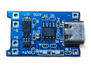
TP4056 Charger Module
Introducing our TP4056 charger module, a compact and efficient solution for charging lithium-ion and lithium polymer batteries in a wide range of electronic applications. This module is designed to provide reliable charging capabilities while ensuring safety and ease of use, making it ideal for DIY projects, prototyping, and portable device applications.
SKU: tp4056-charger-module -
Soldering Items
Soldering Iron 25 Watt
A 25-watt soldering iron is ideal for electronics repair, assembly, and DIY projects. It provides precise control with a temperature range of 250°C to 400°C. Lightweight and ergonomic, it features interchangeable tips for versatile use, ensuring accurate and clean solder joints for delicate components and small-scale tasks.
SKU: soldering-iron -
Sensors and Modules
Soil Moisture Sensor Module
The Soil Moisture Sensor measures the volumetric water content in soil, providing crucial data for agriculture and gardening applications. It typically features two probes that detect soil moisture levels and output a corresponding signal to a microcontroller. This sensor helps optimize irrigation, prevent overwatering, and monitor soil health, making it ideal for automated watering systems and environmental monitoring projects.
SKU: soil-moisture-sensor -
Robot Parts
Motor Driver L298N
Efficient and versatile Motor Driver L298N for controlling DC motors and stepper motors. Ideal for robotics and automation projects, it supports dual H-Bridge configuration and delivers reliable performance. Compatible with Arduino and other microcontrollers, perfect for DIY electronics enthusiasts and professionals seeking precise motor control solutions.
SKU: motor-driver-l298n